The Role of Mucosa In The Nasal Cavity
The entire inner cavities of the nose: airway cavities + all three pairs of turbinates + the sinus cavities, on both sides, are covered with a thick layer of tissue rich with blood vessels and central nervous system nerve endings, called – Nasal Mucosal Tissue.
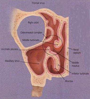
Fig 9: Right side nasal cavities and sinuses.
The respiratory epithelium layer supports the cilia – which is a fine exterior layer of what looks like a blanket of tiny fine hairs, which move in a wave-like formation – that is known as the “cilia-beat”. The cilia “beats” around 250 times a minute and is responsible for capturing the potentially harmful airborne particles (dust, germs and viruses, etc’) allowing them to be trapped in the mucous secreted from the mucous producing glands (the “goblet” and “serum” cells located in the epithelium layer). The debris is pushed towards the nasopharynx and swallowed through the eusophgus into the stomach. It takes the healthy cilia around 20 minutes to push the trapped particle/organism from the anterior nares to the nasopharynx). On a normal day the nasal mucosa will produce about ¾ of a liter of mucous, that kills the invading microorganisms, enables the cilia to push the debris away smoothly, and humidifies the inspired air.

Fig 10: Mucosa Layers of right inferior turbinate cross section (under microscope).
|
Fig 11(a): cross section of nasal mucosal layer. Notice that the cilia is beating. |
Fig 11(b): cross section of respiratory epithelium (drawing). |
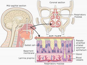
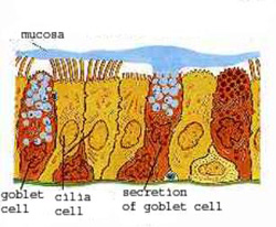
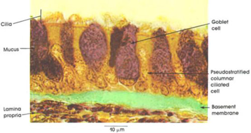
Fig 12: cross section of pseudostratified respiratory epithelium (under microscope).
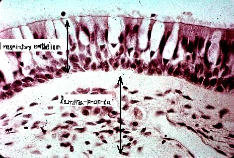
Fig 13: Nasal respiratory epithelium and supporting erectile mucosa (long magnification under microscope).